|
|
|
|
|
|
Page 2 |
Fluorescein Angiogram / Color Pictures |
The following photos were taken with a Topcon TRC-50ia digital ophthalmic/retina camera. |
* This site is BEST viewed at 800 X 600 screen resolution. If the pictures below appear "grainy" or "jagged" it may be due to your video card (this is common with older video cards) or your display settings. But you should still get the idea anyway. |
The following pictures were taken by and are the property of the web designer and may not be reproduced without written permission of the web designer. |
Please be patient while the images download, Thank you! |
|
|
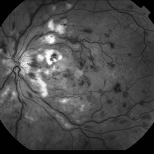
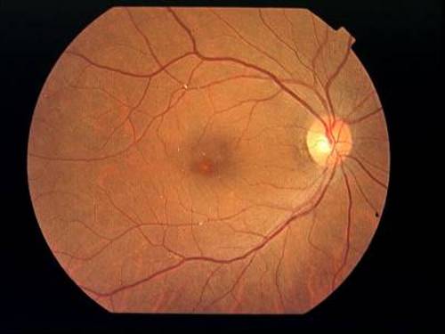
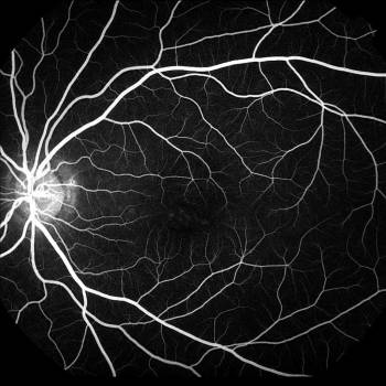
Branch Retinal Vein Occlusion, Right eye before laser treatment. |
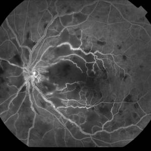
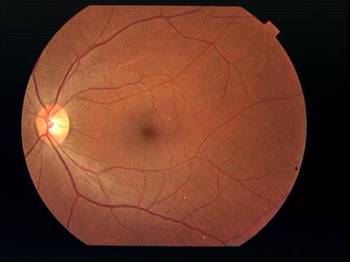
Branch Retinal Vein Occlusion, Right eye after laser treatment. |
|
|
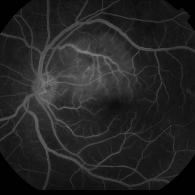
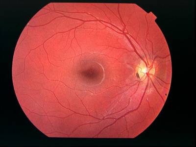
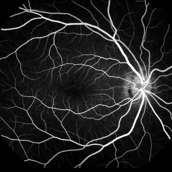
Central Retinal Vein Occlusion, Left eye, "Red-Free" picture. |
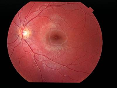
Central Retinal Vein Occlusion, Left eye, "FA" picture. |
|
|
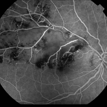
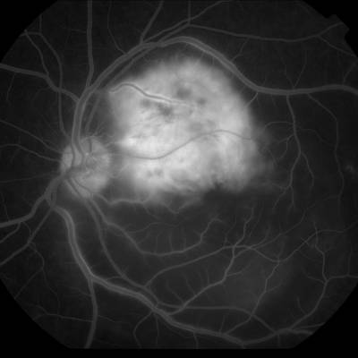
Lesion, Left eye, "Early FA" picture. |
|
|
|
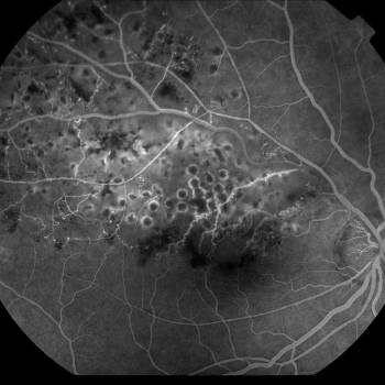
Same Lesion, Left eye, "Mid FA" picture. |
|
|
|
Plaque (Little white spots), Right eye. |
|
|
|
Plaque (Little white spots), Left eye. |
|
|
|
Stargardts Disease, Right eye, "Color picture" |
Stargardts Disease, Left eye, "Color picture" |
|
|
Stargardts Disease, Right eye, "FA picture" |
Stargardts Disease, Left eye, "FA picture" |
|
All of the photos on this site were taken by David Bronson at : |
|
|
Use the menu below for further site navigation: |
Main | Photo gallery, page 1 | Triangle NC Area HospitalsAbout the Designer | Pharmacy List |
|